ISSUES
Generation of Hair-Bearing Skin Organoids from Human Pluripotent Stem Cells by Dr. Jin Kim
(Stem Cell Therapy & Organoids Session 1)
Generation of Hair-Bearing Skin Organoids from Human Pluripotent Stem Cells
Jin Kim_Postdoctoral Researcher, Harvard Medical School
Dr. Jin Kim talked about the logic and method for generating hair bearing skin organoid model starting from Human pluripotent stem cells. She briefly discussed other in vitro cell culture systems which has been used for a long time and are still being used for cell expansion due to efficiency and reproducibility. Dr. Kim also elaborated that organ function comes from complex interactions between different cell populations and not from a single cell type. She then mentioned that in earlier times, when the stem cell technology was still new, the usage of mature somatic cells isolated from tissues was common. She continued to explain that even after stem cell engineering emerged, these cells are still favorable as they are easier to characterize than stem cell derived cells. However, due to insufficient cell sources especially in case of human cells and maybe the co-culture condition, it has not become feasible. For that reason, people started using stem cells since the key feature of stem cell is self-renewal and due to improved stem cell medium, stem cells can be easily expanded. Dr. Kim gave an emphasis that there are adult stem cells with multipotency and pluripotent stem cells that can differentiate into all cell types. By differentiating stem cells, multiple cell types with self-oriented 3D structures can be available.
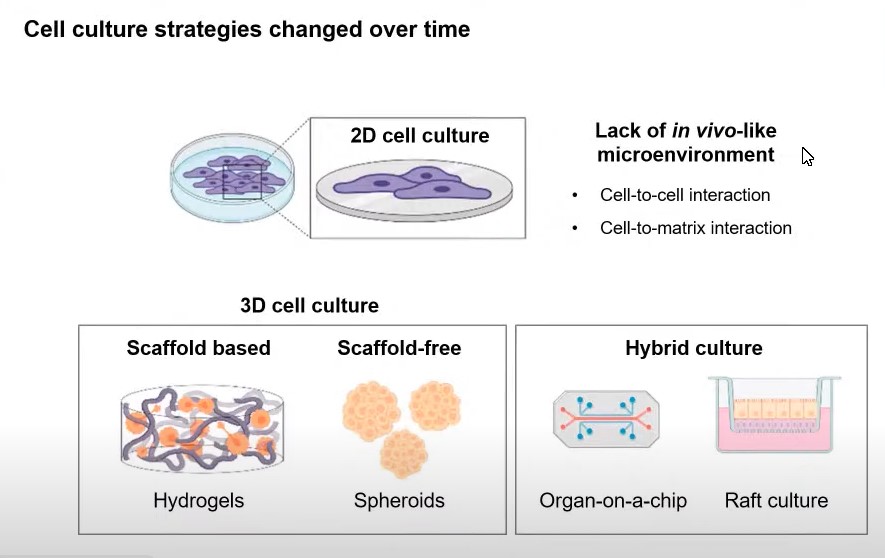
Image Sources: Zoom Presentation Screenshot by Dr. Jin Kim
Dr. Kim further explained that KOEHLER LAB has been using fully potent stem cells to produce hair bearing skin organized by co-inducing ectoderm and mesoderm lineages to form hair and other appendages in skin organoids. Dr. Kim showed the usage of pluripotent stem cells following embryonic development to a certain degree with some images and live video clips. The skin organoid development showed similar features of neurulation forming surface ectoderm on the surface then differentiating mesenchymal cells migrate and cover the outer epithelial layer forming a cyst. According to Dr. Kim’s presentation, like in Vivo neurulation, neural crest cells differentiated in this stage later forming neurons. Outer mesenchymal layer becomes dermis and epithelium differentiate into epidermis and their interactions form hair follicle growing upward. After the video clips were shown from specific days of development, Dr. Kim continued to explain types of skin and hair which their lab produces. She mentioned types of skin in the chin, cheek, and outer ear based on the Single Cell RNA sequencing data. As skin organoids are transplanted into the mouse skin the organoid hair shaft outgrowth and sebaceous gland are seen with cornified layer and later rich like structures comparable to adult facial skin. Dr. Kim explained skin organoid can be used for a wide range of skin focused research from a basic developmental biology to translational projects like the early development of human skin hair follicle and diverse sensory neurons free from ethical issues or drug screening targeting hair growth. Skin organoid can be used for disease modeling like skin cancers, infection, chemicals using patient-derived IPS cells to model genetic disorders. Lastly, she continued to explain that skin organoid should be a cell source for skin reconstructions and wound healing process.
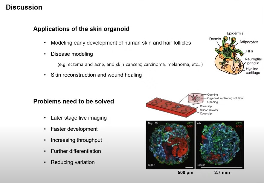
Image Sources: Zoom Presentation Screenshot by Dr. Jin Kim
After giving information on the usage of skin organoids, she made it clear that even though the skin organoid model has a huge potential, it has problems too. Live Imaging is critical for in vitro screening tools and our model is good in the early time points but not the later stage because of its huge size and thick layers. In addition, image-based quantification is also difficult due to the large size and polarized structure. Dr. Kim pointed out that KOEHLER LAB has been trying to clear organoids and image both sides by using silicon isolator but it is a time and resource consuming process to scale up the screening and it takes about 60 days to see hair packs and the end stage takes more than 140 days only representing second trimester stage of fetal skin. Dr. Kim emphasize the need to speed up the differentiation while finding a way to further much more this model and the reduction of variations. On the final part of Dr. Kim’s talk, she gave an update that she is now trying hard to address these issues by further optimizing signaling cues during the early induction stages. Dr. Kim is mainly working on organoid on a chip approach for better Imaging and quantification throughout the long-term culture. She ended her talk with the hopes that KOEHLER LAB can share the improved version of their model in the future.